Hydrocele
What is Hydrocele?
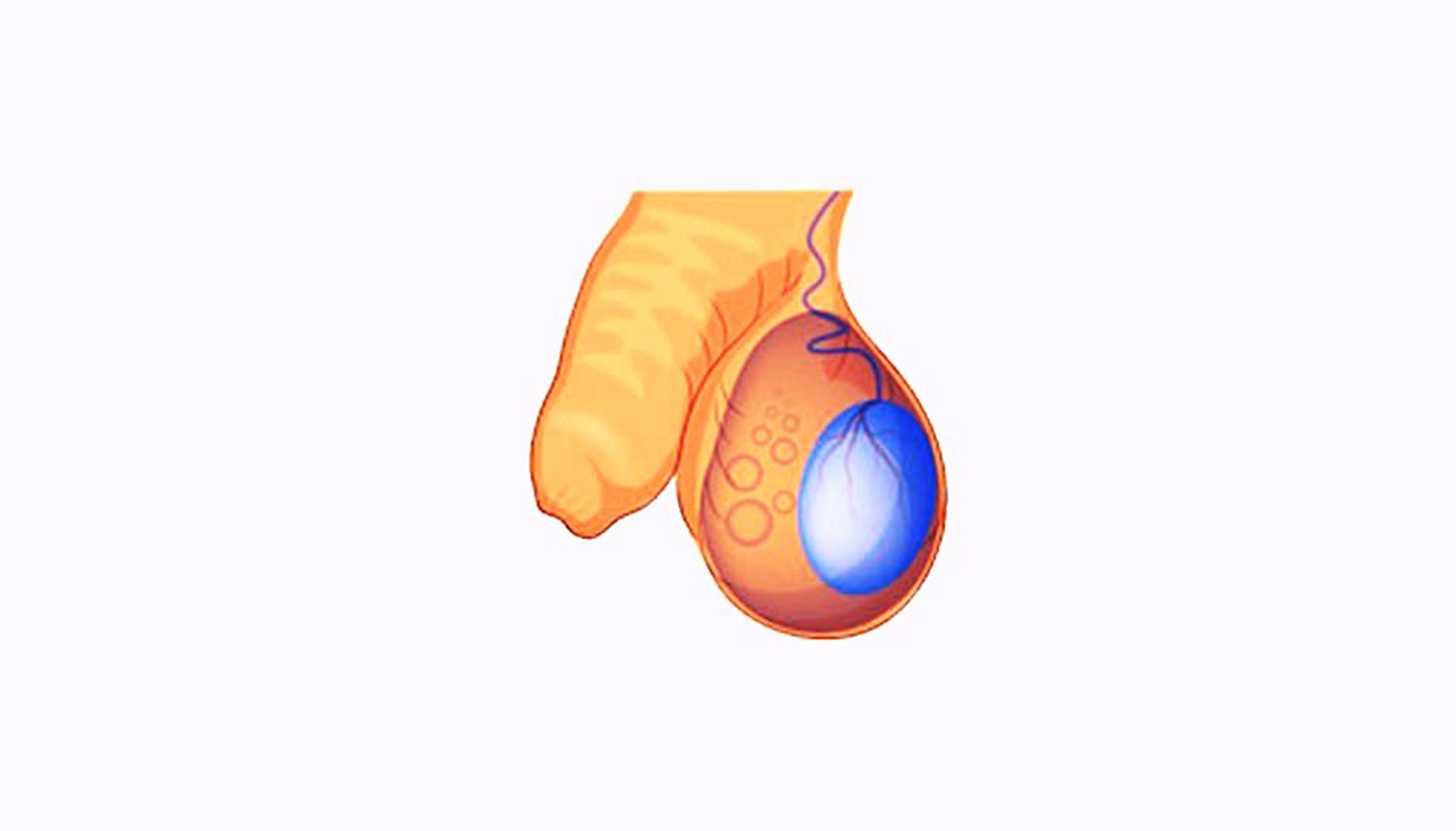
Hydrocele is a condition in which the testicle becomes severely swollen, with more fluid than normal collecting between the membranes surrounding the testicle. Normally, there is 0.5-1.0 ml of fluid in this range to ensure lubrication of the testicle. In hydrocele, this amount of fluid is 200-300 ml and sometimes even more.
Diagnosis
Diagnosis is very easy because the appearance and patient history are very typical. The scrotum appears unilaterally or bilaterally, extremely swollen and tense. It is filled with liquid.
During the examination, a typical feeling of elasticity is felt when touched with a finger.
When you look at the scrotum with a flashlight in a dark room, it turns pink. This simple examination, called the TRANSILLUMINATION sign, proves that there is fluid in this swelling and is TYPICAL for HYDROCELE.
Simple Hydrocele
Hydroceles that are unilateral, do not grow or shrink, are painless, can reach very large sizes, have an unknown cause, are tense, and have an oval shape are called simple hydroceles.
Secondary Symptomatic Hydrocele
It usually occurs in adulthood as a result of inflammation or tumors in the testicles or related structures inside the bags. Chronic hydrocele may develop in 10-15% of testicular tumors. For this reason, in suspected cases, it should be checked whether there is any other accompanying disorder with scrotal ultrasonography.
**The only treatment for hydrocele is surgery.
Hydrocelectomy
Temporarily inserting a needle and draining the fluid inside (PERCUTANEOUS ASPIRATION) is a method that should not be used as it tends to recur in a short time and will cause adhesions in the future operation.
The picture above shows the hydrocele sac that was released from the scrotum and removed from the incision line during the surgery. During the surgery, this sac is opened, the water is drained, the sac leaves are turned upside down and stitched, and the testicle is placed in the scrotum.
Spermatocele / Epididymis Cyst
They are small, painless masses located above and behind the testicle. It is a cystic formation containing dead sperm.
A cystic structure occurs as a result of the accumulation of sperm. The reason is unknown. It does not cause pain. The patient notices a hardness or swelling inside the scrotum, separate from the testicle, on the upper back side of the testicle.
Diagnosis is made by examination and ultrasound. If it does not reach large volumes, there is no need for treatment. If it grows too large, it is removed surgically.